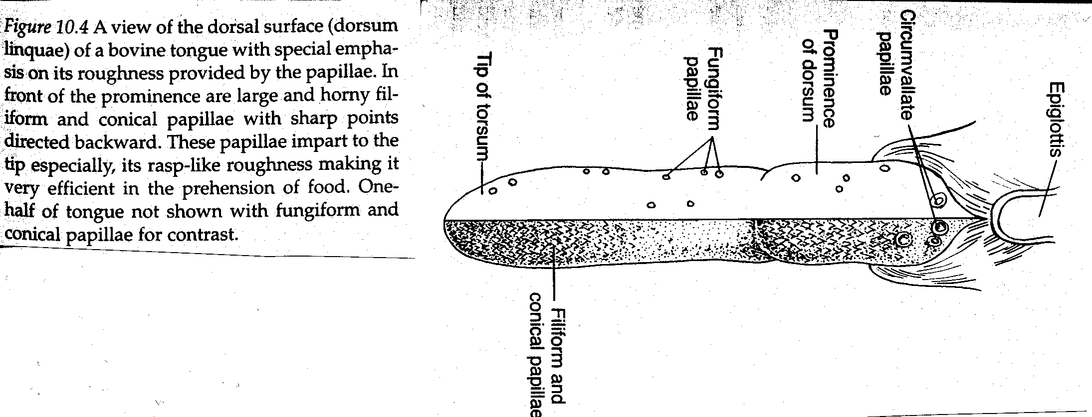
- Tongue
- Muscular organ used to maneuvar the food mass
- Tongue can be differentiated from other muscle tissues because it has fibers oriented in three directions.
- Rough surface of the tongue is provided by numerous projections known as papillae
- Provide traction for moving the food within the mouth
- Help in grooming themselves and offspring
- Process of digestion is aided by discriminatory taste buds
Located on the tongue surface within the vallate and fungiform papillae
- Pharynx
- Common passageway for food and air
- Opens into mouth , nasal cavities, Eustachian tubes, larynx and esophagus.
- During passage of food through pharynx it is prevented from entering larynx and nasal cavities because of reflex and mechanical factors.
- Esophagus
- Muscular tube extending from the pharynx to the stomach
- During its course to the stomach the esophagus traverses the thorax within the mediastinal space.

- Esophagus then passes through the diaphragm into abdominal
Cavity and enters the stomach
- Food and fluid are moved from the pharynx to the stomach by
Muscular waves of contraction.
- The esophagus is normally closed at the pharyngeal end by
Tonic activity of the cranio-esophageal sphincter.
- The esophagus remains closed at the opening to the stomach
(cardia) because of a closure that is physiologic in nature.
- On its way to the stomach, the esophagus courses along the left
Side of the trachea. Bolus transport can be observed by watching the left side of the neck (especially in ruminants)
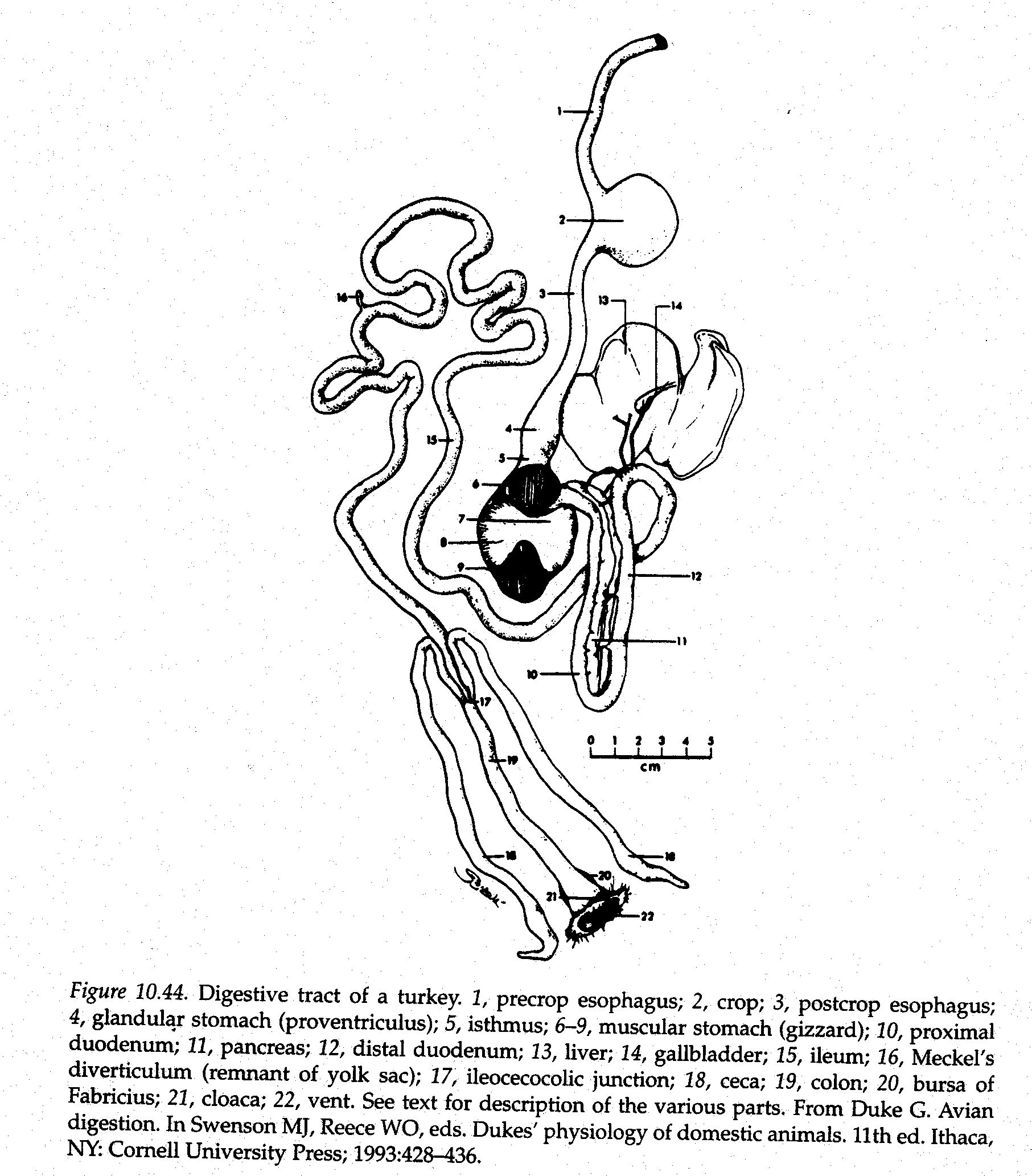
- Stomach
- Food is received by the stomach for storage (pending further
Digestion) and for continuation of digestion.
- Primary regions as viewed from outside:
- Cardia
- Located nearest the esophagus
- Continued by the fundus
- Fundus
- Dome-shaped part of the stomach
- Fundus is adjacent to the Corpus
- Corpus
- Rounded base or bottome
- Antrum
- Constricted part that joins duodenum
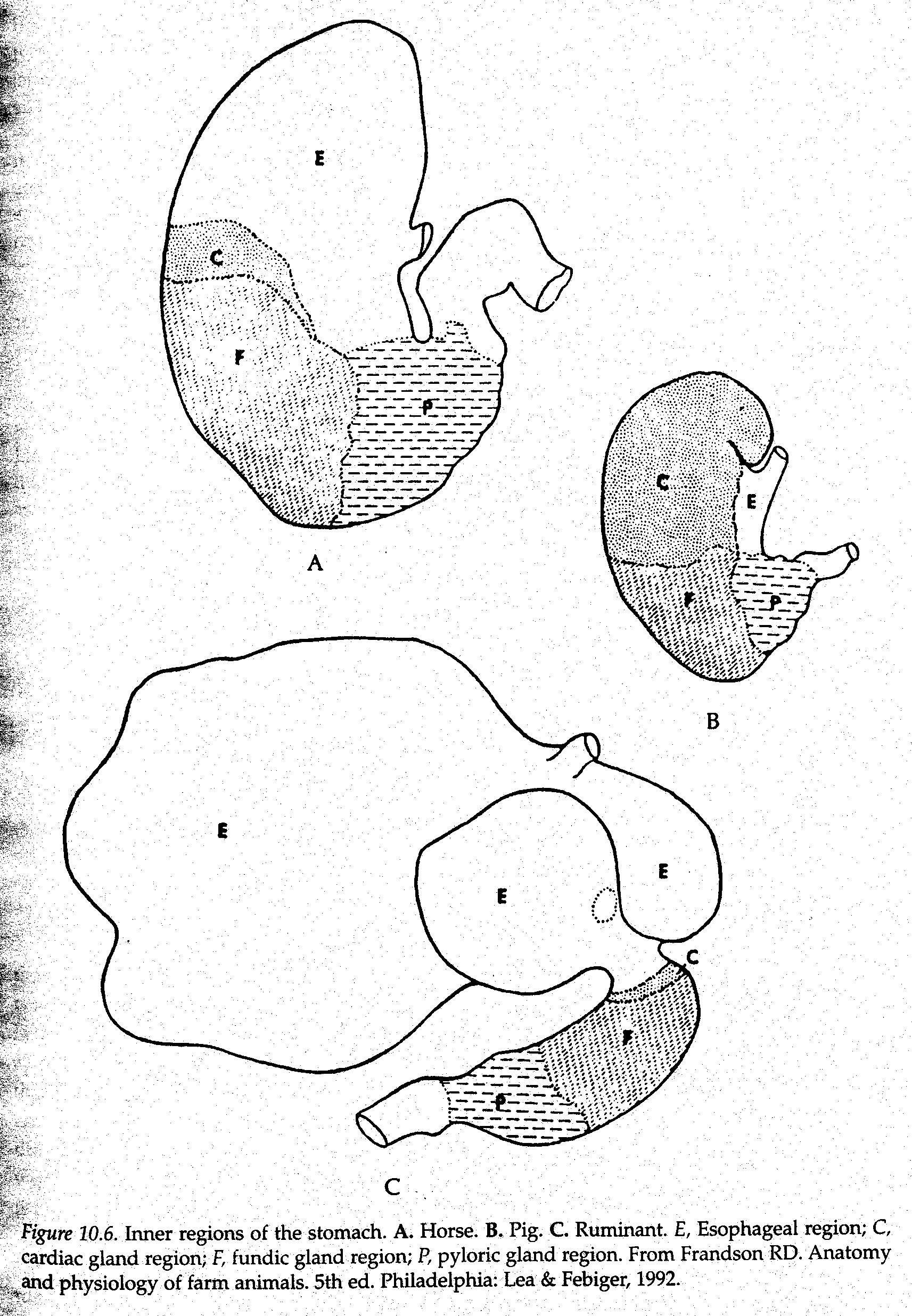
- Regions of stomach as defined by cell type (inner aspect)
- Esophageal gland region
- cardiac gland region
- Secretes mucous
- fundic gland region
- entire space between cardiac and pyloric gland regions.
- These glands sometimes called gastric glands
- Secrete hydrochloric acid and pepsinogen
- pyloric gland region
- secrete mucous and the hormone gastrin
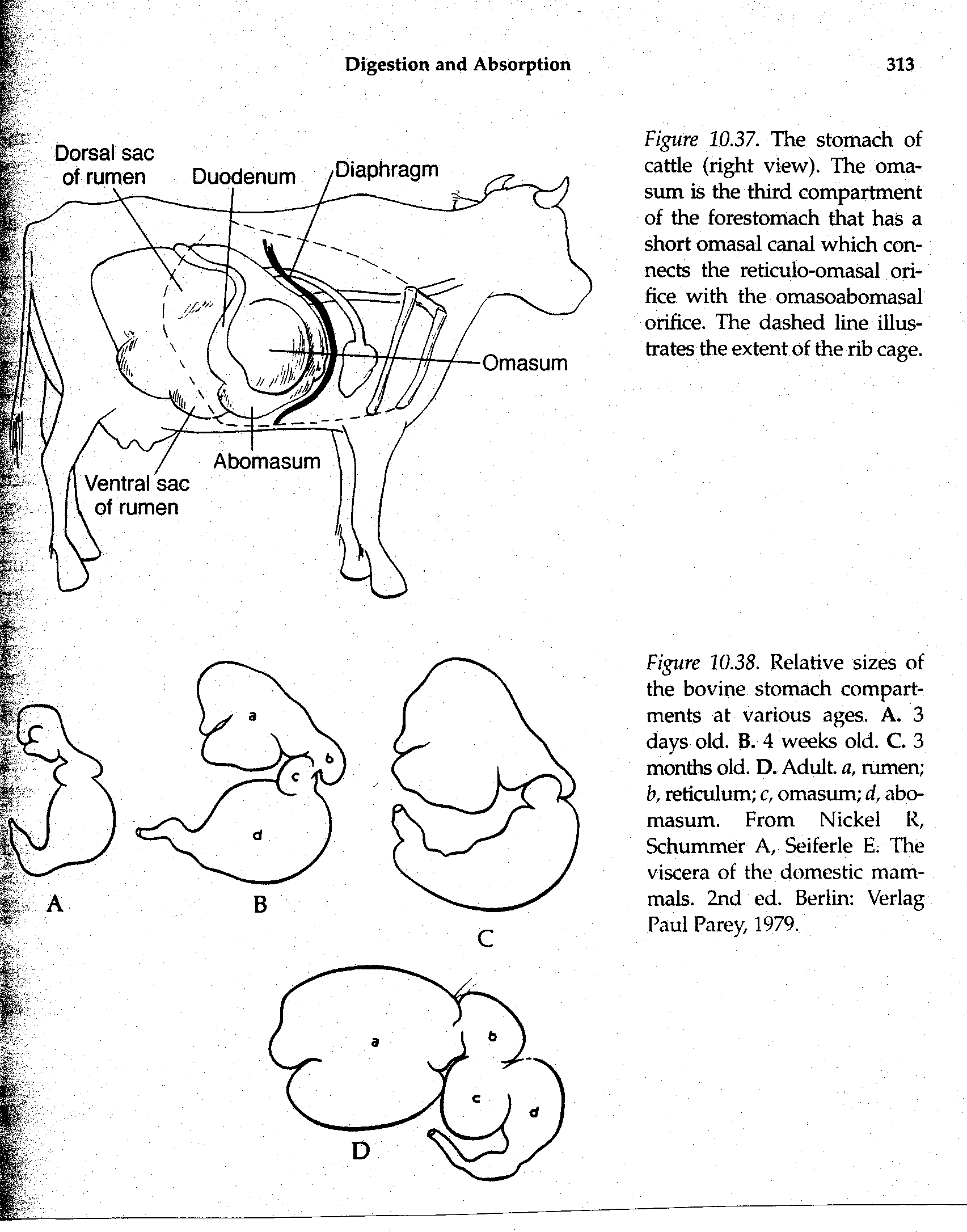
- Ruminant Stomachs
- Foregut fermentation
- Includes the rumen, reticulum (omasum) and abomasum
- True stomach is the abomasums
- Small Intestine
- Stomach contents enter the small intestine after their preparation in the stomach.
- Small intestine comprised of three segments
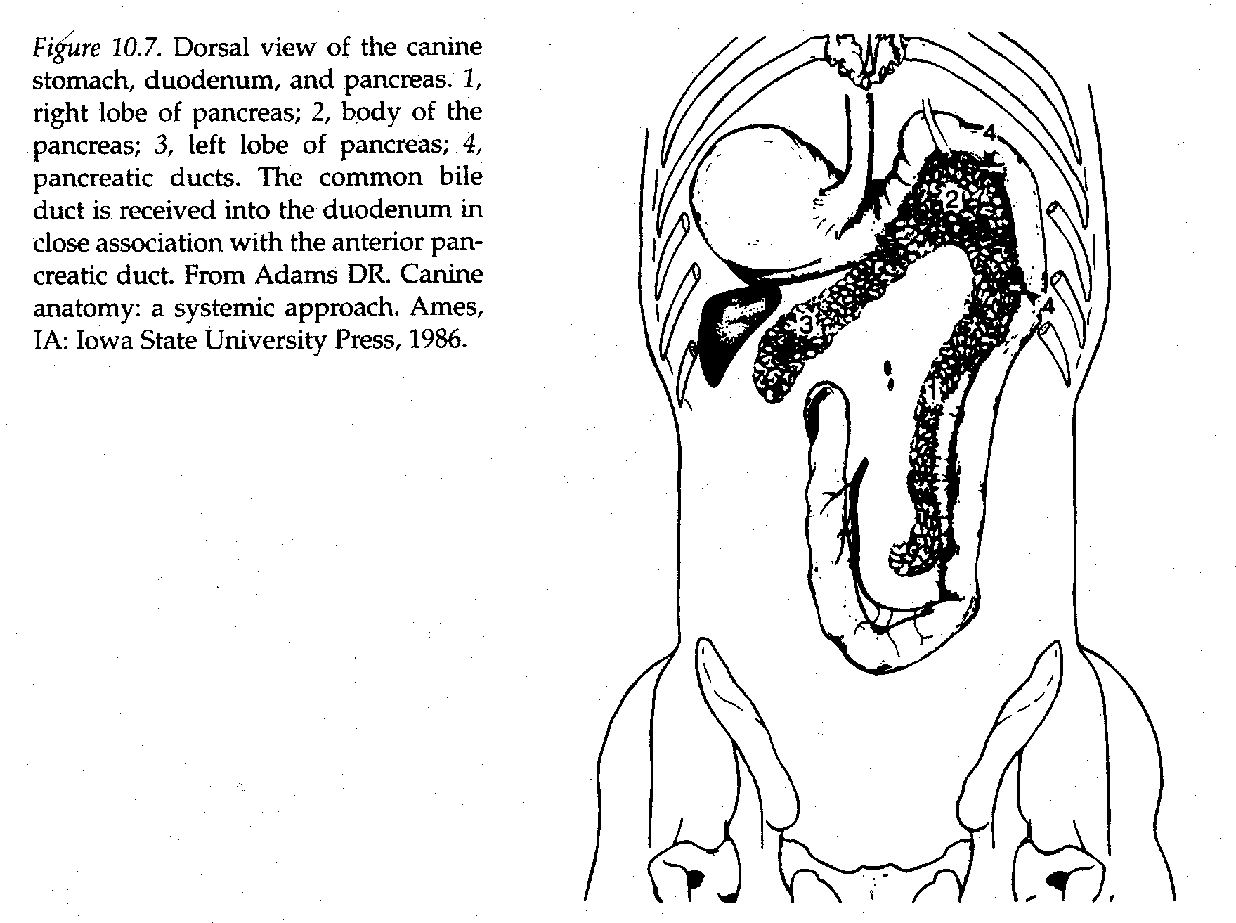
- duodenum
- closely connected to the pancreas
- receives pancreatic secretions
- Also receives bile
- jejunum
- ilium
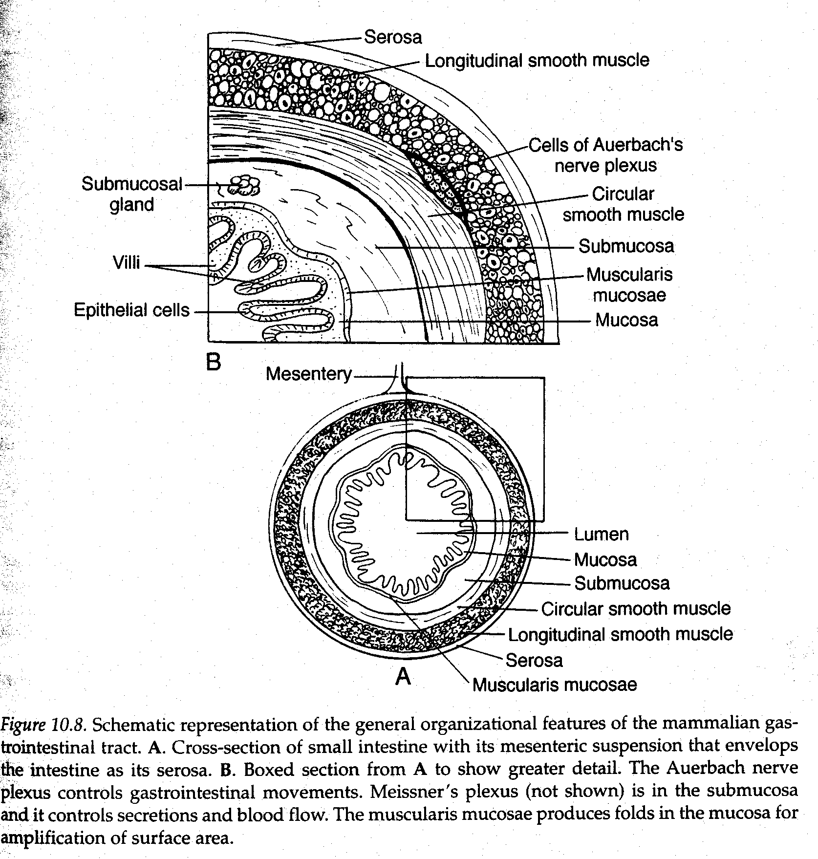
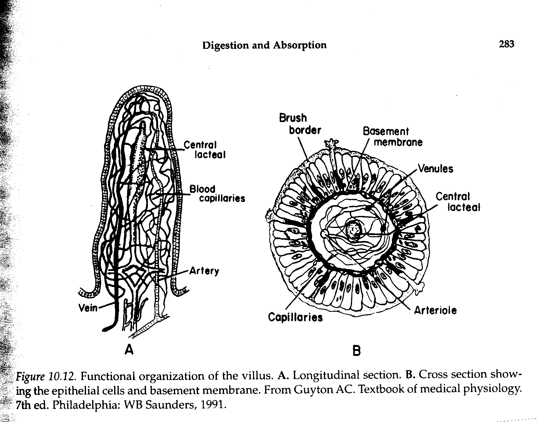
- Inner layer of the small intestine having intimate contact with the contents of the lumen is composed of an epithelial cell layer known as the mucosa.
- The submucosa is a connective tissue layer that provides space for blood vessels, lymph vessels and nerve fibers.
- There is also a sparse layer of smooth muscle fibers known as the muscularis mucosa
- Individual fibers from muscularis mucosa attach to villi and cause movement when contracting.
- A nerve network (Meissner’s plexus) in the submucosa is important in controlling secretions of the epithelial cells and blood flow.
- This network also serves a sensory function via stretch receptors
- Another nerve plexus (Auerbach’s plexus), between the inner circular and outer longitudinal muscle layers is important in controlling gastrointestinal movement.
- These two nerve plexus’s are referred to as the enteric nervous system and it extends from the esophagus to the anus.
- Outer layer of the intestine is the serosa. It covers the
Intestine and is continuous with the mesentery which suspends the intestine within the abdominal cavity.
- The mesentery is in turn continuous with the
Peritoneum.
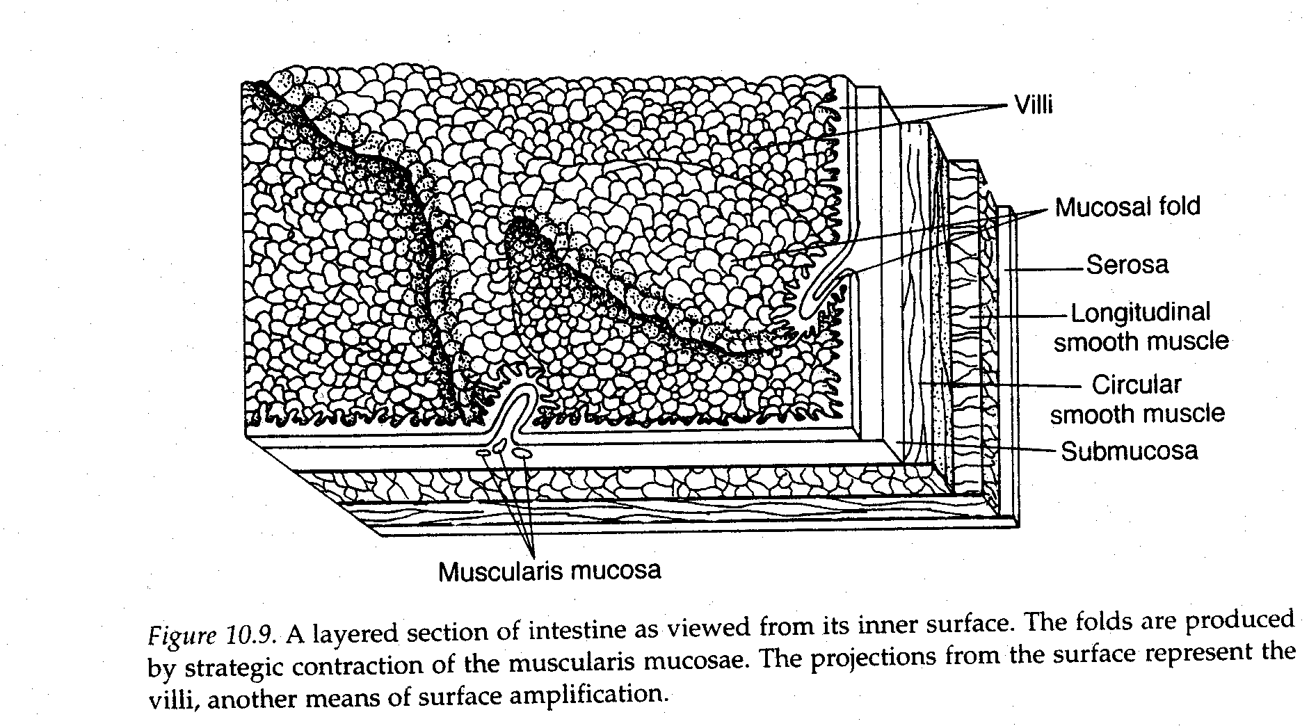
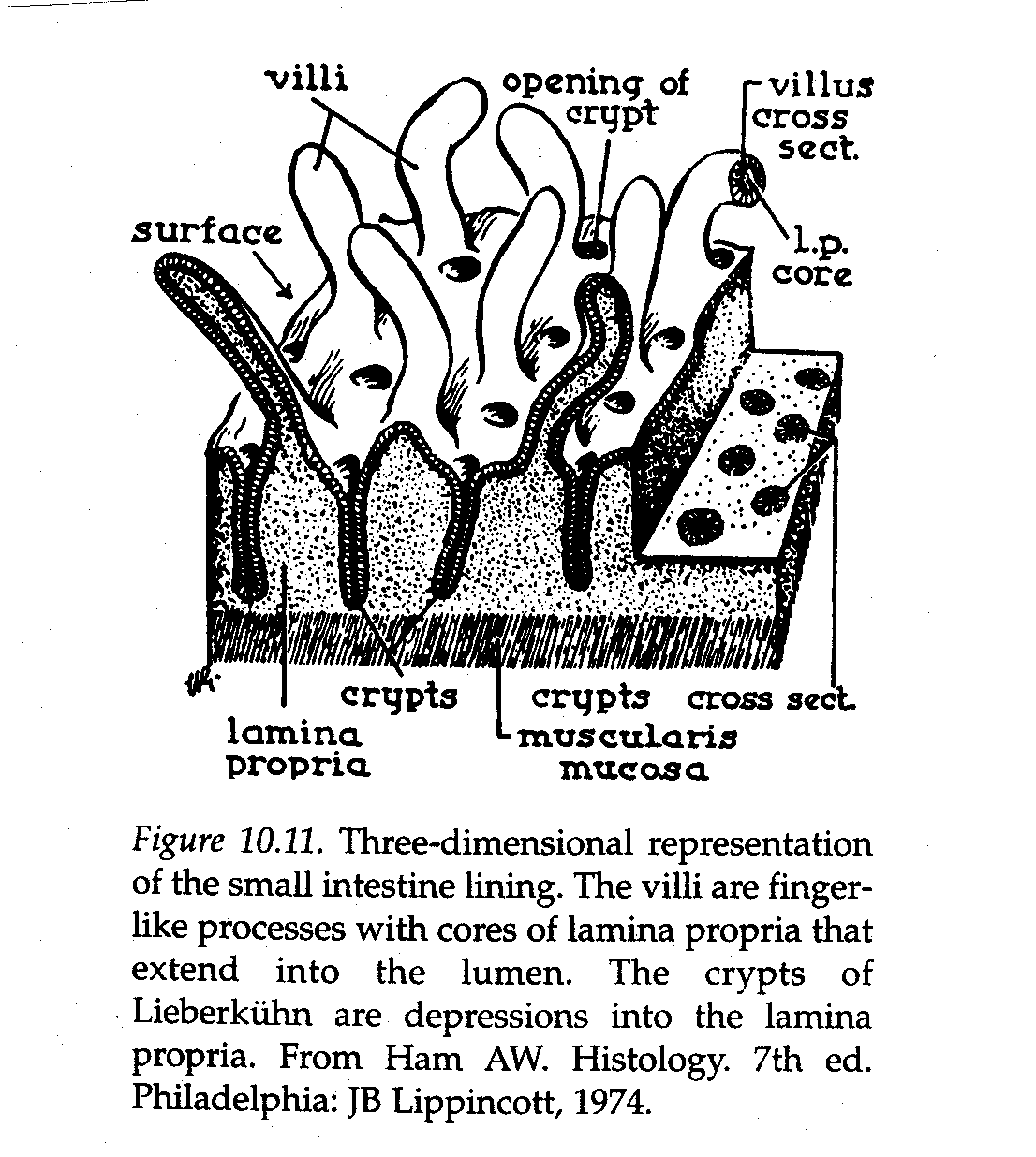
- The surface area of the intestine is increased by long length and folding of the tissue within the intestine.
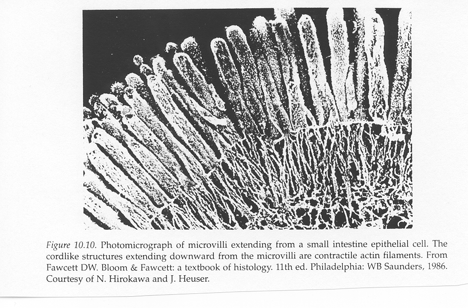
- Folds or placations are covered with villi and the individual cells that cover the villi have their own microvilli.
- Microvilli provide for the greatest amplification of surface area and constitute the brush border.
- This amplification increases the total surface area of the small intestine about 600 times that of
A smooth cylinder of the same size.
- The crypts of liberkÜ
hn are cloistered groups of
Undifferentiated cells between adjacent villi.
- Only cells of villi to undergo division
- Renewal of cells in the villi is accomplished by migration of new cells away from the crypts.
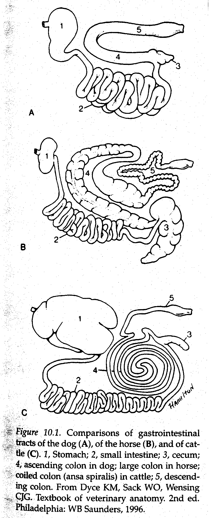
- Large Intestine
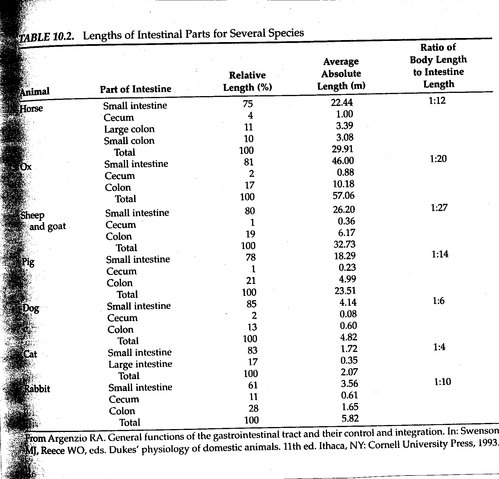
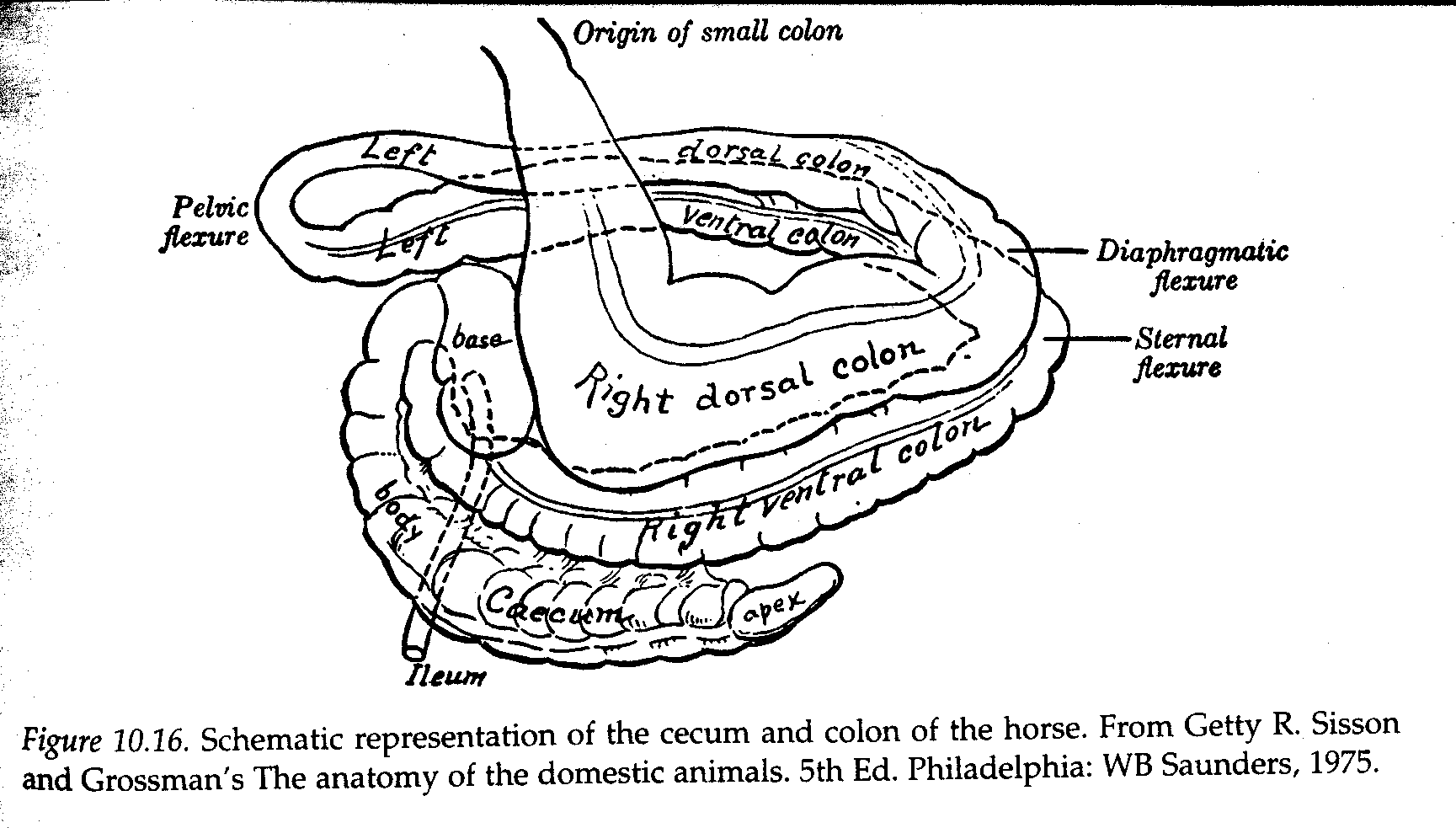
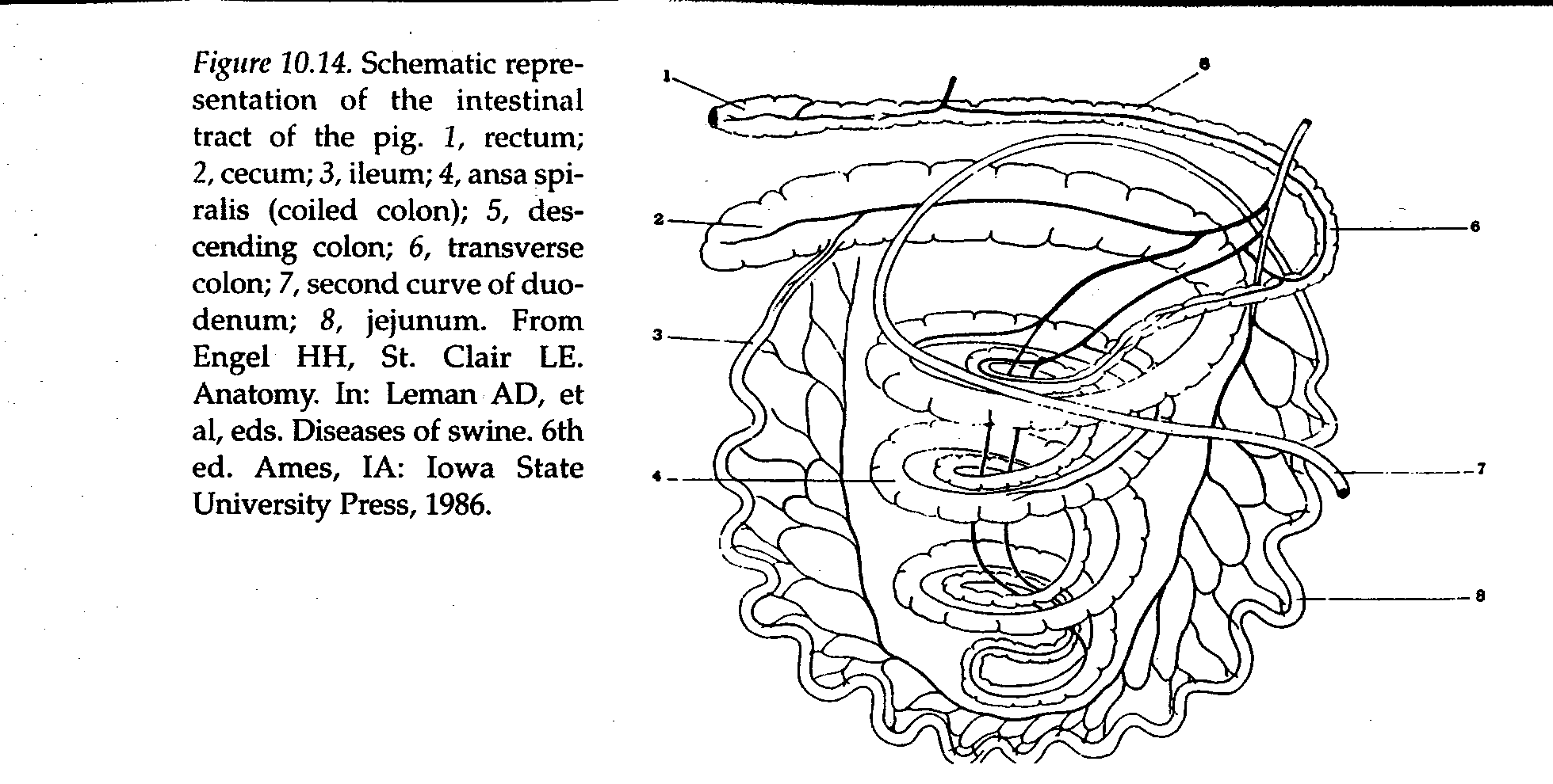
- Contents from the terminal part of the ileum enter the large intestine at the cecum (ileocecal junction) in the horse, at the colon(ileocolic junction) in the dog or at the cecumand colon (ileocecolcolic junction) in the ruminant and pig.
- The large intestine consists of the cecum and colon.
- Development of the large intestine varies among animals according to diet.
- In ruminants bacterial and protozoal cells are digested here.
- In simple herbivores, (e.g. horse) enzymatic digestion preceeds fermentation and no microbes or protozoa are available for digestion here.
- Food requiring further digestion by fermentation enters or is diverted into the cecum unless it is developed poorly as in the dog.
- The colon continues from the cecum to its termination at the
Anus.


- Accessory Glands
- Salivary Glands
- Three pairs of well-defined glands
- Glands connected to mouth by ducts
- Salivary glands are serous, mucous or mixed depending on secretion
- Blood vessels and nerves enter each gland where ducts exit
- Innervation is sympathetic and parasympathetic

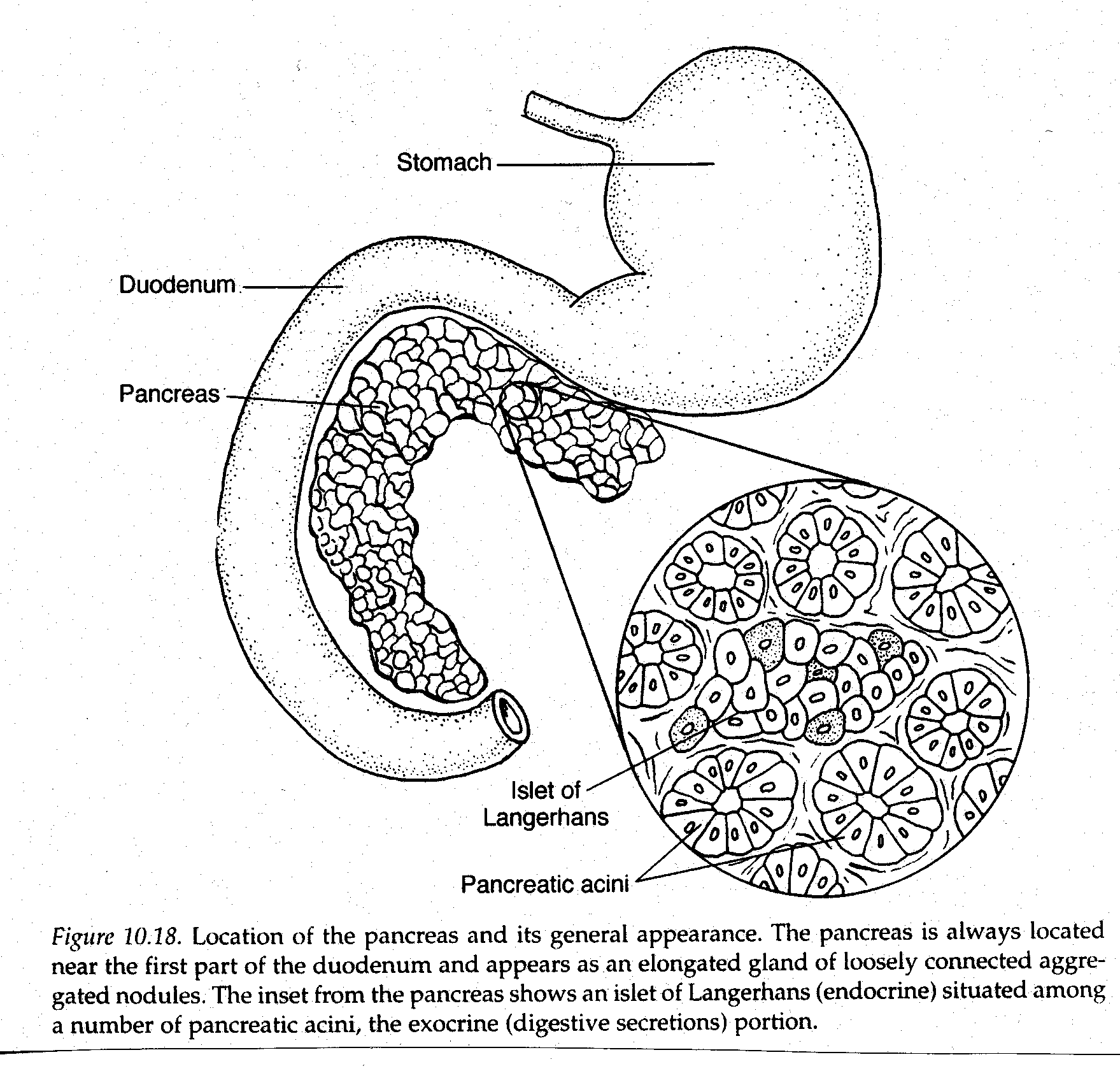
- Pancreas
- has both endocrine and exocrine function
- Always located near the first part of the duodenum
- appears as elongated gland of loosely connected nodules
- Main pancreatic duct enters duodenum near common bile duct.
- In sheep and goats pancreatic duct enters bile duct
- Endocrine cells are scattered throughout pancreas
- Islets of langerhans
- Alpha cells produce glucagons
- Beta cells produce insulin
- Cells secrete directly into the bloodstream
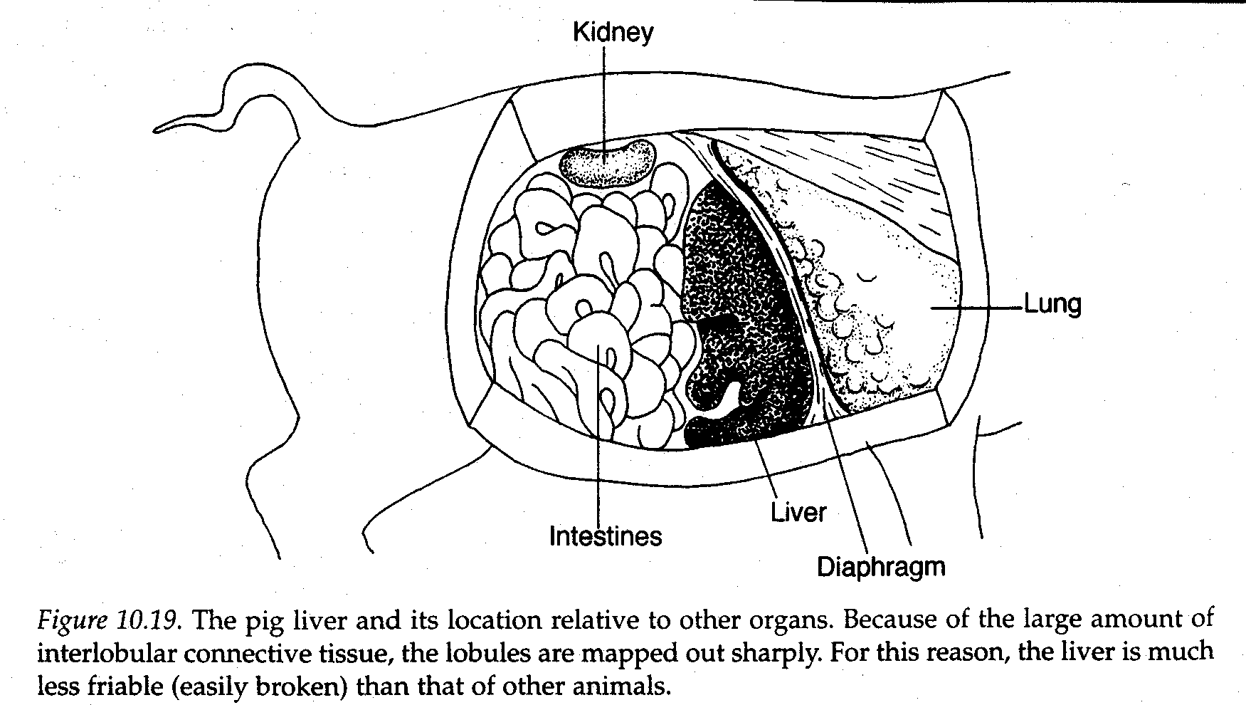
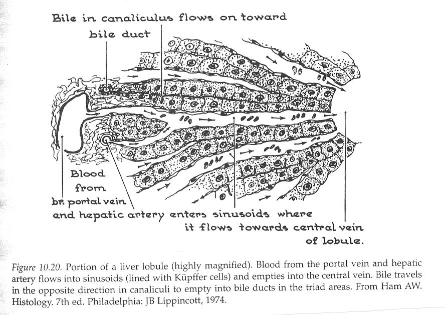
- Liver
- Multipurpose organ
- Always located immediately behind the diaphragm
- In ruminants it tends to be on the right side
- Lobules of the liver are clearly demarcated
- Liver receives arterial blood through hepatic artery and venous blood through portal vein.
- Portal vein drains stomach, spleen, pancreas and intestines.
- Blood from both sources is circulated through the sinusoids
- Here it is detoxified and modified before returning via the hepatic vein to the vena cava